Orthopedics
Orthopedic Surgery
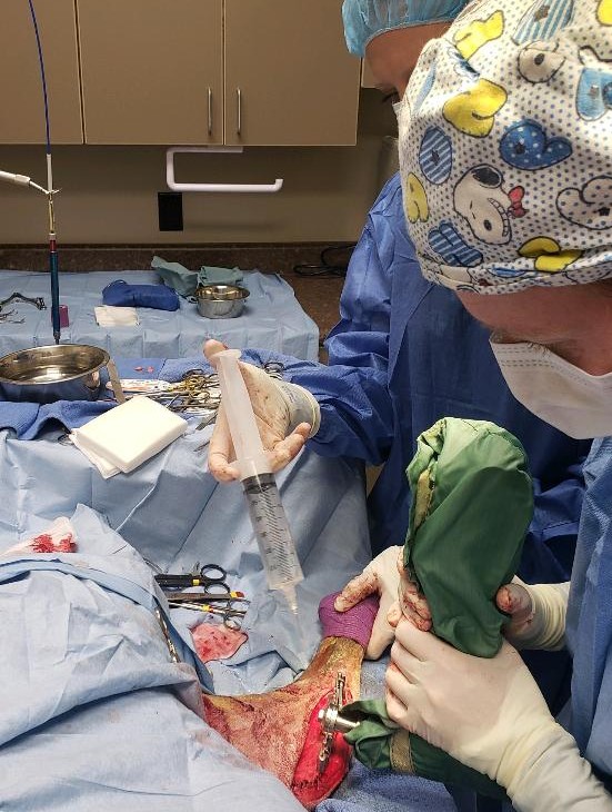
Orthopedic surgery is any surgery that involves the skeletal system. Orthopedic surgery aims to improve your pet’s quality of life by easing pain, re-establishing function and stability, and bettering your pet’s range of motion.
At Continental Animal Wellness Center we are proud to offer our patients orthopedic surgery using a state of the art surgical laser that helps to improve the surgical outcome.

Common Orthopedic Surgeries Offered at Continental:

Femoral Head Ostectomy (FHO)
An FHO, or femoral head ostectomy, is a surgical procedure that aims to restore pain-free mobility to a diseased or damaged hip by removing the head and neck of the femur (the long leg bone or thighbone).
When the hip becomes damaged or diseased, however, this mobility can be affected. If the acetabulum and the head of the femur do not fit together properly, this poor fit can influence the degree of movement that the joint can achieve. In addition, this poor joint fit can lead to chronic pain and inflammation.
An FHO restores mobility to the hip by removing the head of the femur. This removes the ball of the ball-and-socket joint, leaving just an empty socket. The muscles of the leg will initially hold the femur in place and, over time, scar tissue will form between the acetabulum and the femur to provide cushioning that is referred to as a ‘false joint’. Although this joint is anatomically very different from a normal hip joint, it provides pain-free mobility in most patients.
There are four main reasons why an FHO might be performed on your pet: fractures involving the hip, luxations/ subluxations of the hip joint, chronic severe arthritis, and avascular necrosis of the femoral head.

Limb/ Digit Amputations
An amputation is a surgical procedure where a pet’s limb is removed. An amputation may be performed due to a variety of reasons which may include a cancer diagnosis, severe trauma, fractures, infections, or chronic pain.
Typical post-operative recovery from an amputation is approximately two weeks until the site is healed. Dogs and cats carry slightly more weight on the forelimbs than the hindlimbs, so recovery from a hindlimb amputation may not require as much assistance during recovery. Complications or mobility issues may prolong recovery. Infrequent complications may include those related to anesthesia, incisional irritation or infection, bleeding, or fluid build-up at the surgical site.
Long-term, patient quality of life is great following an amputation. Most amputees have no long-term limitations or pain. Additional rehabilitation therapy can be helpful in the transition phase after surgery to strengthen other limb muscles. Amputees frequently run, jog with their owners, swim, and jump on and off the couch with ease. It is important once your pet has had a limb amputation that they are maintained in ideal body condition, given joint supplements and is encouraged to exercise daily. Dogs and cats can do quite well on 3 limbs, however, after an amputation, it can be quite challenging if your pet injures another limb in the future.
Toe/ digit amputations are also performed for similar reasons where the removal of a single digit is all that is necessary to accomplish the end result.
Resources:
purina.co.uk
acvs.org

Extracapsular Lateral Suture Stabilization
The lateral suture method involves placing a nylon material on the outside of the knee in a similar orientation to the injured cranial cruciate ligament. This stabilizes the joint and allows fibrous tissue to develop which provides long-term stabilization after the nylon degrades. The majority of dogs recover back to 80-85% of normal within 6 months and may improve further by 1 year. However, smaller dogs are better candidates for this procedure compared to larger dogs, active dogs, or working dogs
Cruciate Ligament Disease
Cranial cruciate ligament disease is the most commonly diagnosed orthopedic condition in canines. Any breed or age of dog can rupture their cranial cruciate ligament but in general cruciate disease is more commonly seen in large-breed dogs. Unlike traumatic ACL injuries in humans, dogs typically have a degenerative injury to the ligament with a very high rate of the injury happening in both knees. There are many surgical options available to stabilize the knee after a cruciate ligament rupture. Depending on activity level, size of the patient, intended level of exercise, and budget, one of the many surgeries can be selected by your veterinarian to best suit your pet’s needs.
Hip Dysplasia
Hip dysplasia is a deformity of the hip joint which happens during growth. The hip is a ball and socket joint and during growth both the ball and socket must grow at the same rate. When the growth does not occur at equal rates it results in laxity of the joint and subsequent arthritis and degenerative joint disease. Hip dysplasia can occur in all types of dogs but most commonly affects large-breed dogs and it is a genetic disease that seems to be affected by diet, environment, exercise, and growth rate. Hip dysplasia is diagnosed through a sedated orthopedic exam combined with radiographs. Treatment includes diet, supplements, physical therapy, cold laser therapy, and anti-inflammatories. Over time the laxity in the joint leads to increasing arthritis and pain and a lack of mobility for your pet. Occasionally an FHO may be recommended to help your pet increase mobility and decrease pain.
